15 Feb, 2024
MRI Scan for Cancer Detection: How Does It Work and When Is It Used?
An MRI scan is a common method used in the diagnosis, assessment and treatment of many different types of cancer. It can be used to determine whether a tumour is cancerous or not, and helps doctors to understand whether cancer has spread (metastasized).
MRI can also show whether treatment has been effective, and if any cancerous tissues remain after treatment.
The scanning process is painless and doesn't use any radiation, so it will not cause harm if you require multiple scans throughout treatment and aftercare.
What is cancer?
Cancer is a condition where abnormal body cells grow and divide uncontrollably. This means they can invade surrounding healthy tissues, and destroy the tissues. There are more than 200 types of cancer, across different organs and body parts.
Uncontrollable growth and multiplication of cancer cells can be caused by genetic changes in the cells, and can result in a tumour or lump.
Sometimes, cancer that begins in one part of the body can spread to another part, which is called metastasis. The first cancer is called the primary, and secondary cancers occur through metastasis.
Doctors can grade cancer into different stages, based on how big it is, and if it has spread or not. These stages give an idea of the treatments that might work best, and how quickly the cancer could grow.
Why do I need an MRI scan for cancer?
If you have discovered a lump in your body, or are suffering from symptoms of cancer such as unexplained bleeding, unexpected weight loss, or changes to your bodily functions, an MRI scan is a key imaging method to either diagnose or rule out cancer.
MRI scans are useful for:
-
locating tumours that aren’t visible externally
-
deciding whether a tumour is or isn’t cancerous
-
finding out how big a tumour is
-
seeing whether cancer has spread
-
measuring blood flow in the area of the tumour
-
checking whether treatment is working, for example radiotherapy or chemotherapy
-
making sure cancer hasn’t come back in the months and years following treatment (in remission)
Dealing with the prospect of cancer can be a worrying time for you and your loved ones. It is important to get a diagnosis as soon as possible, and have any lumps or concerns seen by your doctor or examined using MRI.
Early diagnosis is the key to a better prognosis, and optimises the opportunities for treatment.
How accurate is Magnetic Resonance Imaging in detecting cancer?
Research has shown that MRI scans are 77% accurate when distinguishing between malignant (cancerous) and benign (non-cancerous) tumours. This is one reason why it is the preferred modality for imaging and evaluating soft-tissue tumours.
MRI scans also find nearly [90%](https://www.radiologytoday.net/archive/rt0915p7A.shtml#:~:text=Around 90%25 of all breast,detection rate of just 37.5%25.) of all breast cancers - more than ultrasounds and mammograms combined. MRIs are usually more successful at finding tumours than they are at distinguishing between benign and malignant.
Alongside accuracy, there are other important measures to ensure the success of a diagnostic test:
-
sensitivity is a measure of how often a test is correct when a positive result is generated for people who do have a condition
-
specificity measures how well a test correctly generates a negative result for people who don’t have a condition
MRI scans have a specificity of 85% when differentiating between non-cancerous and cancerous tumours, which means false negatives are low.
What does cancer look like on an MRI?
MRI scan images are usually black and white, with various shades of grey. MRIs also produce images from different angles (cross sectional images) all around the body, and are best at showing soft tissues.
Cancerous tissue can show up on MRI images as a white or very light mass, whereas it would be dark in colour on an ultrasound image. Contrast dye, which is a substance injected into the body before some MRI scans, enables the cancer to present more brightly on MRI scan images. They can cause you to experience a metallic taste and warming sensation during your scan, but it's important to let your radiographer know if you have previously had an allergic reaction to a contrast medium such as gadolinium.
Can an MRI detect cancer anywhere in the body?
MRI scans are best for imaging soft tissues, including the brain, nerves, organs, cartilage, tendons, muscles and ligaments. They can generate very detailed images that can be used for diagnosing cancer. They can also be used to stage cancer, which means finding out how advanced it may be, or how early it has been found.
Therefore, MRI scans are particularly good for detecting:
-
brain tumours
-
primary bone tumours
-
soft tissue sarcomas in muscle, tendons, fat, blood vessels and nerves.
-
tumours affecting the spinal cord
-
some forms of bone marrow cancer
-
tumours in the pelvic organs and reproductive system (prostate cancer, bladder cancer, uterine cancer, ovarian cancer and certain liver cancers)
-
Recent research even showed that MRI screening was a more effective method of identifying prostate cancer than previous physical DRE (digital rectal examination) or PSA blood tests.
Sometimes, your imaging test will form part of a series of tests including blood tests, biopsies and various scans.
MRI scan vs CT scan for cancer: what other imaging tests are there?
If you have metal fragments, implants, shrapnel, medical devices like cochlear implants and surgical clips, or other permanent metal objects implanted in your body, you may be recommended a CT scan instead of an MRI scan.
This will depend on the type of cancer investigation needed, as CT is not always suitable for soft tissue imaging, but the MRI machine's strong magnetic field can interact with metal objects, body piercings, artificial joints and other metal items.
CT scans for cancer
When choosing an MRI or CT scan for cancer detection, it's important to speak to medical professionals, as cancer is a complex health issue that benefits from early detection. There are also some limitations and safety concerns for each scan type, such as metal objects being affected by MRI's magnetic field, while CT scans use ionising radiation in the form of X-rays, that can be harmful if over-used.
Getting the right scan quickly is key for fast diagnosis and access to treatment.
The key differences between MRI and CT for cancer are:
-
Lung imaging - CT is the modality of choice for spotting and staging lung cancer
-
CT also can be used to check the whole body for metastases (cancer cells that have spread elsewhere in the body) quickly and efficiently
-
CT is also commonly used for staging cancers
-
CT is not as good for identifying cancer in the pelvic organs - MRI would usually be chosen in these cases, such as for the prostate, uterus, and certain liver cancers.
Compared to MRI, it's important to consider: how accurate is ct scanning for cancer? It really depends on the type of cancer the scan is aimed to examine. The lungs and colon are most commonly scanned with CT for cancer detection, while MRI is preferred for the soft tissues and some pelvic organs, as well as brain cancer imaging.
How does MRI work?
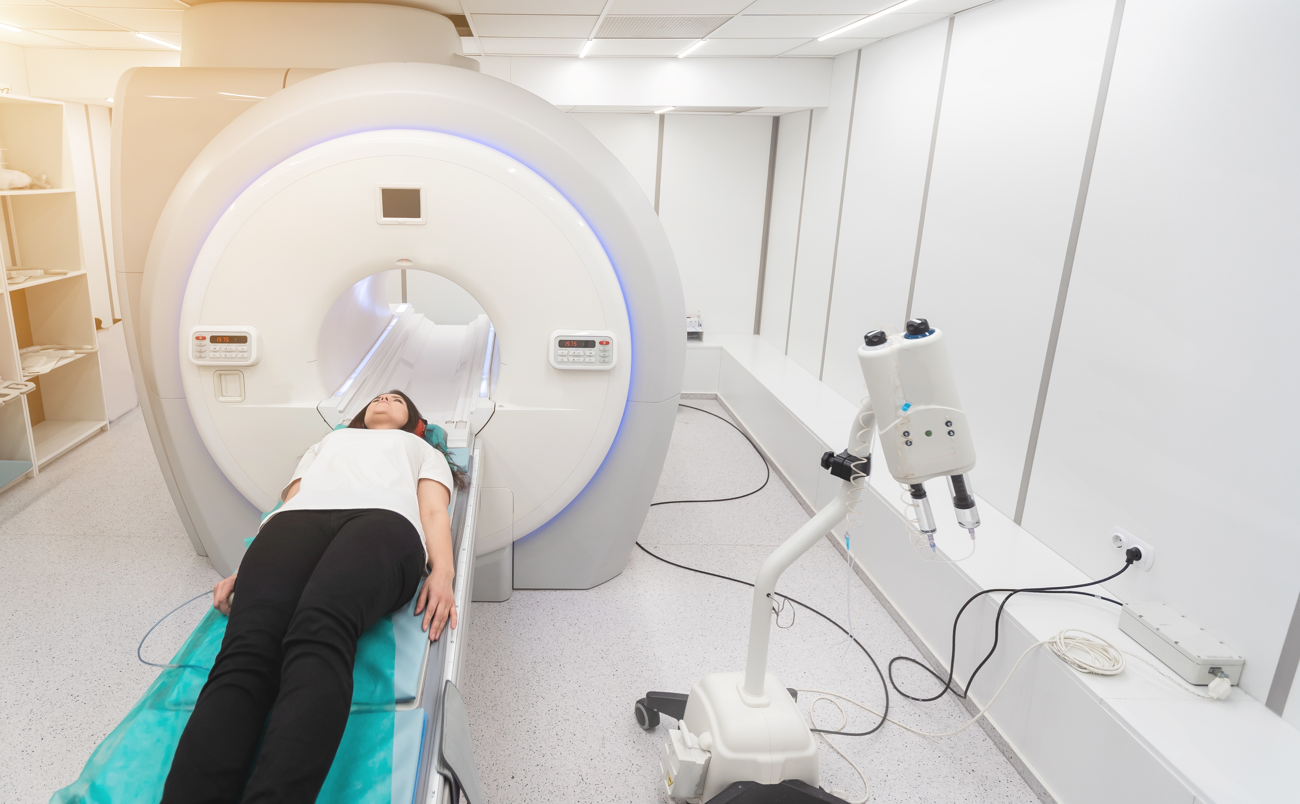
MRI machines use strong magnets and radio waves to create detailed pictures of the inside of your body, without any risks associated with ionising radiation. An standard MRI machine looks like a short tunnel, but some types of MRI scanner are open with magnets above and below you. A flat table moves in and out of the scanner, and you lie very still on it during your imaging test.
MRI is usually done at an outpatient imaging centre or hospital department.
Sources used:
Cancer Research UK. (2022, October 27). What is cancer? https://www.cancerresearchuk.org/about-cancer/what-is-cancer
Cancer Research UK. (2023, October 10). Stages of cancer. https://www.cancerresearchuk.org/about-cancer/what-is-cancer/stages-of-cancer
CT Scan vs. MRI: What’s the Difference? And How Do Doctors Choose. (2022, December 8). Memorial Sloan Kettering Cancer Center. https://www.mskcc.org/news/ct-vs-mri-what-s-difference-and-how-do-doctors-choose-which-imaging-method-use
England, N. (n.d.). NHS England » Earlier diagnosis. https://www.england.nhs.uk/cancer/early-diagnosis/
MRI scan. (n.d.). Tests and Scans | Cancer Research UK. https://www.cancerresearchuk.org/about-cancer/cancer-in-general/tests/mri-scan
RAGCP. (n.d.). Clinical guidance for MRI referral [PDF]. https://www.racgp.org.au/download/Documents/Guidelines/MRI referrals/clinicalguidancemrireferral.pdf
Soft tissue Sarcoma—Patient version. (n.d.). National Cancer Institute. https://www.cancer.gov/types/soft-tissue-sarcoma
Study: MRI has best breast cancer detection rate. (n.d.). https://www.radiologytoday.net/archive/rt0915p7A.shtml
UCSF Department of Radiology & Biomedical Imaging. (2016, February 6). What does cancer look like? UCSF Radiology. https://radiology.ucsf.edu/patient-care/for-patients/video/what-does-cancer-looks
Website, N. (2024, February 2). Cancer. nhs.uk. https://www.nhs.uk/conditions/cancer/
Wj, C., Chung, H., Mj, S., Sh, L., Mh, L., Js, L., Kim, D. Y., & Lee, W. (2012). MRI to differentiate benign from malignant soft-tissue tumours of the extremities: a simplified systematic imaging approach using depth, size and heterogeneity of signal intensity. British Journal of Radiology, 85(1018), e831–e836. https://doi.org/10.1259/bjr/27487871
Tips for understanding studies. (n.d.). Health News Review. https://www.healthnewsreview.org/toolkit/tips-for-understanding-studies/understanding-medical-tests-sensitivity-specificity-and-positive-predictive-value/